By Mark S. Parker
The ebook is wonderfully illustrated with up to date radiographs, 64-MDCT CT scans, and multiplanar CT, CT
angiographic and a few MR and 3-D imaging. greater than 1,500 fine quality pictures make the examining effortless and pleasant.
The captions are concise and critical. The textual content is phenomenal and straightforward. -- Pediatric Radiology
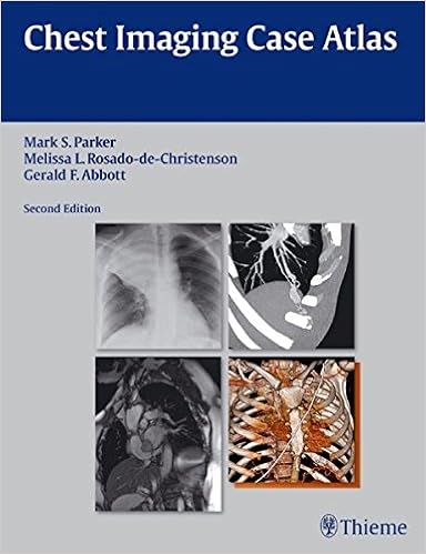
Written by means of well known specialists in chest imaging, Chest Imaging Case Atlas, moment Edition allows radiology citizens, fellows, and practitioners to hone their diagnostic abilities by way of instructing them the right way to interpret plenty of radiologic circumstances. This atlas comprises over two hundred instances on stipulations starting from Adenoid Cystic Carcinoma to Wegener Granulomatosis. every one case is supported through a dialogue of the affliction, its underlying pathology, normal and strange imaging findings, administration, and analysis, supplying a entire review of every disorder.
Special beneficial properties of the second one Edition:
- Over 1500 high quality pictures demonstrating common and pathologic findings and their adaptations
- More multiplanar, CT angiographic (CTA), MRI, and 3D imaging is integrated into the textual content, aiding readers remain present with this speedily altering expertise
- 40 new situations and up to date photos in instances from the earlier variation
- A new post-thoracotomy chest part addresses general post-operative findings and problems linked to universal thoracic interventional systems
- The neoplastic illnesses part contains the recent TNM staging procedure for lung melanoma
- The grownup heart problems part now includes a dialogue on univentricular and biventricular or end-stage center failure together with a variety of ventricular help units and the complete synthetic middle, their imaging good points, and problems linked to their use
- The diffuse lung ailment part has been extended to contain an method of HRCT interpretation
- Case discussions are in keeping with up-to-datereviews of present literature in addition to vintage landmark articles
- Pearls are supplied to explain the good points which may strongly help a particular prognosis, permitting readers to sharpen their scientific diagnostic skills
This publication is a useful illustrated reference that every one physicians in radiology and chest imaging specifically, together with pulmonary medication physicians and thoracic surgeons, must have on their bookshelf.
Read Online or Download Chest Imaging Case Atlas PDF
Similar pulmonary & thoracic medicine books
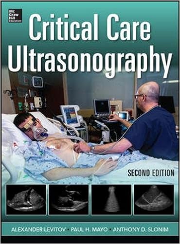
An entire, hands-on advisor to profitable photograph acquisition and interpretation on the bedside ''The genuine power of this textbook is its medical concentration. The editors are to be complimented on preserving a constant constitution inside of each one bankruptcy, starting with simple actual rules, sensible “knobology,” scanning tips, key findings, pitfalls and barriers, and the way the foremost findings relate to bedside patho-physiology and decision-making.
This factor brilliantly pairs a rheumatologist with a pulmonologist to discover all the 14 article topics. subject matters contain autoantibody checking out, ultility of bronchoalveolar lavage in autoimmune illness, and pulmonary manifestations of such stipulations as scleroderma, rheumatoid arthritis, lupus erythematosus, Sjogren's Syndrome, Inflammatory Myopathies, and Relapsing Polychondritis.
Comparative Biology of the Normal Lung, Second Edition
Comparative Biology of the conventional Lung, 2d variation, deals a rigorous and complete reference for all these concerned about pulmonary learn. This absolutely up-to-date paintings is split into sections on anatomy and morphology, body structure, biochemistry, and immunological reaction. It maintains to supply a special comparative viewpoint at the mammalian lung.
Notice what workout trying out can exhibit approximately cardiopulmonary, vascular, and muscular well-being. Now in its 5th Edition, Principles of workout trying out and Interpretation continues to bring well timed details at the body structure and pathophysiology of workout and their relevance to medical drugs.
- Respiratory System
- Medical devices for respiratory dysfunction : principles and modeling of continuous positive airway pressure (CPAP)
- Managing Breathlessness in Clinical Practice
- Environmental and Occupational Medicine
- Environmental and Occupational Medicine
Additional info for Chest Imaging Case Atlas
Example text
20â•… Pulmonary venous anatomy—conventional pulmonary venous anatomy. (A) Non-gated MDCT (RPO epicardial view) shows single right and left superior and inferior pulmonary veins draining into the left atrium without accessory veins. LI, left inferior pulmonary vein; LS, left superior pulmonary vein; RI, right inferior pulmonary vein; RS, right superior pulmonary vein. (B) Common (conjoined) veins. Volume rendered CT (PA epicardial view) from a non-gated MDCT shows a right common pulmonary vein (RCV).
40) is the interface formed by the contact between the left upper lobe and the mediastinal reflection between the aortic arch superiorly and the left pulmonary artery inferiorly (Fig. 40B). It is characteristically either straight or concave laterally. On cross-sectional imaging, the aortopulmonary window refers to the mediastinal space bound laterally by the left parietal pleura and medially by the ligamentum arteriosum. 38â•… Section Iâ•… Normal Thoracic Anatomy A Fig. 38â•… Radiologic depiction of the posterior junction line.
22G) This fissure separates the anterior segment of the right upper lobe from the middle lobe. It is seen in 44–88% of normal chest radiographs (Figs. 22B). In two-thirds of studies it lies at the fourth anterior intercostal space. On MDCT, it is most often recognized as an oval lucent area devoid of vasculature (Fig. 22C). On 8% of studies, the fissure is seen as an oval area of ground glass attenuation. Coronal and sagittal MIP reconstructions clearly delineate the course and orientation of the horizontal fissure relative to the other standard fissures (Figs.


