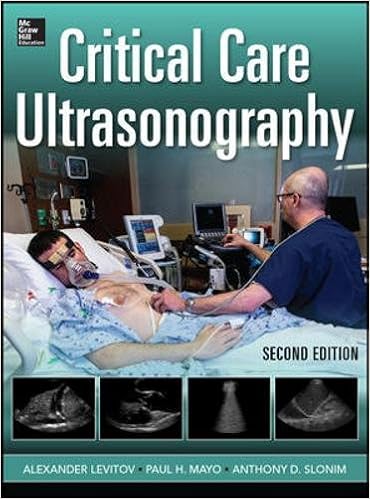
By Alexander Levitov, Paul Mayo, Anthony Slonim
A whole, hands-on advisor to profitable photo acquisition and interpretation on the bedside
''The genuine energy of this textbook is its scientific concentration. The editors are to be complimented on protecting a constant constitution inside of each one bankruptcy, starting with easy actual rules, sensible “knobology,” scanning suggestions, key findings, pitfalls and barriers, and the way the most important findings relate to bedside patho-physiology and decision-making. therefore, the authors have succeeded in supplying a reference normal to aid bedside severe care services to appreciate the context for his or her own sonographically guided decision-making processes.''-- serious Care medication
Written by means of best practitioners within the box, this all-in-one text/DVD package deal is full of sensible assistance that is helping you grasp medical ultrasonography in a severe care setting. right here, you’ll research precisely tips on how to make the most of diagnostic ultrasound as a part of the actual examination, because the ebook examines present facts helping its use within the significantly sick grownup and child.
Organized by means of physique approach, severe Care Ultrasonography good points self-contained chapters that may be used as person reference publications for a number of interventions, from transthoracic echocardiography to echocardiagraphic review of cardiac trauma. via this in-depth insurance, you’ll get a feeling of the way this crucial expertise helps the cross-disciplinary nature of severe care. The book’s authoritative content material is bolstered all through by way of a full-color presentation and enormous quantities of concept-clarifying illustrations, figures, and images.
Features
- Comprehensive insurance of the basics of ultrasound use in severe care
- Guidance on ultrasound tactics permits practitioners to take advantage of ultrasound for vascular and axial approaches, bettering security and making sure that nationally well-known compliance criteria are upheld
- Cardiac ultrasound chapters assist you investigate and visual display unit the patient’s cardiopulmonary prestige non-invasively
- Chapter on Neck and higher breathing Ultrasound deals an summary of little-known concepts that experience no longer been comprehensively defined in the other resource
- Full-color presentation, with 495 illustrations that emphasize the fundamental abilities required to imagine anatomic constructions and interpret findings
- Companion DVD positive aspects movies that hide pediatric serious care, goal-directed echocardiography within the ICU, echocardiographic evaluate of valve functionality and endocarditis, and more!
Read Online or Download Critical Care Ultrasonography PDF
Similar pulmonary & thoracic medicine books
A whole, hands-on consultant to winning snapshot acquisition and interpretation on the bedside ''The genuine energy of this textbook is its medical concentration. The editors are to be complimented on retaining a constant constitution inside of each one bankruptcy, starting with easy actual rules, useful “knobology,” scanning advice, key findings, pitfalls and boundaries, and the way the major findings relate to bedside patho-physiology and decision-making.
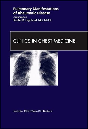
This factor brilliantly pairs a rheumatologist with a pulmonologist to discover all of the 14 article topics. subject matters comprise autoantibody checking out, ultility of bronchoalveolar lavage in autoimmune disorder, and pulmonary manifestations of such stipulations as scleroderma, rheumatoid arthritis, lupus erythematosus, Sjogren's Syndrome, Inflammatory Myopathies, and Relapsing Polychondritis.
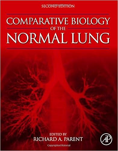
Comparative Biology of the Normal Lung, Second Edition
Comparative Biology of the conventional Lung, 2d variation, deals a rigorous and entire reference for all these keen on pulmonary learn. This totally up-to-date paintings is split into sections on anatomy and morphology, body structure, biochemistry, and immunological reaction. It keeps to supply a different comparative viewpoint at the mammalian lung.
Observe what workout checking out can display approximately cardiopulmonary, vascular, and muscular well-being. Now in its 5th Edition, Principles of workout trying out and Interpretation continues to convey well timed details at the body structure and pathophysiology of workout and their relevance to medical drugs.
- Lung Function Tests Made Easy, 1e
- Pediatric Neurogastroenterology: Gastrointestinal Motility and Functional Disorders in Children
- Air Pollution and Health Effects
- Interventional Pulmonary Medicine
Extra info for Critical Care Ultrasonography
Sample text
The deeper the image, the longer the PRP, the lower the PRF. related in the following ways: PRP (sec) = 1/PRF (Hz) PRF (Hz) = 1/PRP (sec) Note that though it is measured in the same units, PRF has no relationship to the frequency of the ultrasound wave produced during pulse generation that is measured in MHz. 7). Shorter pulses, just as shorter wavelengths of the pulse wave, reflect off the smaller objects. Shorter pulses produce better images by improving axial resolution. Axial resolution can be described by the following relationship: Axial resolution = SPL (mm)/2 DOPPLER PHENOMENON AND ITS USE IN DIAGNOSTIC ULTRASOUND First described by Christian Doppler, the Doppler phenomenon simply states that if the source of sound (transducer) and the object reflecting the sound (reflector) are moving in relationship to each other, the frequency of the reflected sound wave will change.
Related in the following ways: PRP (sec) = 1/PRF (Hz) PRF (Hz) = 1/PRP (sec) Note that though it is measured in the same units, PRF has no relationship to the frequency of the ultrasound wave produced during pulse generation that is measured in MHz. 7). Shorter pulses, just as shorter wavelengths of the pulse wave, reflect off the smaller objects. Shorter pulses produce better images by improving axial resolution. Axial resolution can be described by the following relationship: Axial resolution = SPL (mm)/2 DOPPLER PHENOMENON AND ITS USE IN DIAGNOSTIC ULTRASOUND First described by Christian Doppler, the Doppler phenomenon simply states that if the source of sound (transducer) and the object reflecting the sound (reflector) are moving in relationship to each other, the frequency of the reflected sound wave will change.
In either case, the image quality is degraded. output to the highest safe amplitude, it is incumbent upon the physician–sonographer not to prolong the examination unnecessarily. 3). The returning ultrasound and therefore electrical impulses are very weak and need to be amplified. The amplification, also known as receiver gain, is controlled by the operator and increases amplitude of all signals received by the transducer. In almost all imaging modalities, the amplitude (strength, volume) of the signal is presented on the screen as brightness.


