By Peter Hoskin, Vicky Goh
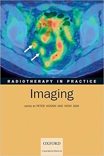
Imaging is a serious part within the supply of radiotherapy to sufferers with malignancy, and this e-book teaches the rules and perform of imaging particular to radiotherapy. Introductory chapters define the elemental rules of the to be had imaging modalities together with x-rays, ultrasound, CT, MR, nuclear drugs, and puppy. website particular chapters then hide the most tumor websites, reviewing optimum imaging innovations for prognosis, staging, radiotherapy making plans, and follow-up for every website. Chapters are co-authored via oncologists and radiologists focusing on a selected quarter to supply an authoritative view at the function of imaging within the patient's trip and examples of appropriate pictures are supplied all through. the $64000 components of radiation safety, publicity justification, and dangers, also are comprehensively coated, exploring concerns equivalent to balancing radiation publicity with long term hazards of radiation results, similar to moment melanoma induction.
Read Online or Download Radiotherapy in Practice - Imaging PDF
Similar radiology & nuclear medicine books
Textbook of Interventional Neurology
Endovascular intervention - utilizing medicine and units brought via catheters or microcatheters put into the blood vessels via a percutaneous strategy - has emerged as a comparatively new minimally invasive method of deal with cerebrovascular sickness and probably intracranial neoplasms. This textbook offers a finished evaluate of ideas pertinent to endovascular remedy of cerebrovascular ailments and intracranial tumors, with a close description of recommendations for those approaches and periprocedural administration techniques.
Radiation Physics with Applications in Medicine and Biology
An creation to nuclear physics such as chapters on current and previous functions of nuclear drugs, nuclear imagery, compartment idea, neutrons and different heavy debris, X-rays and Y-rays. contemporary info at the ideas of radiation safeguard can also be addressed.
A whole introductory textual content to musculoskeletal imaging easy Musculoskeletal Imaging is an engagingly written, accomplished textbook that addresses the elemental ideas and methods of common diagnostic and complicated musculoskeletal imaging. so that it will be as clinically suitable as attainable, the textual content specializes in the stipulations and methods regularly encountered in real-world perform, akin to: top and decrease extremity trauma Axial skeletal trauma Arthritis and an infection Tumors Metabolic bone ailments Bone infarct and osteochondrosis Shoulder, knee, backbone, elbow, wrist, hip, and ankle MRI additionally, you will locate authoritative assurance of: symptoms in musculoskeletal imaging the foremost suggestions of utilizing diverse modalities in musculoskeletal imaging present advances in musculoskeletal scintigraphy The ebook is superior through fantastic figures and illustrations, together with a four-page full-color insert; "Pearls" that summarize must-know info; and a good creation to musculoskeletal ultrasound by means of foreign specialists from France and Brazil.

Esophageal Cancer: Prevention, Diagnosis and Therapy
This publication stories the hot development made within the prevention, analysis, and therapy of esophageal melanoma. Epidemiology, molecular biology, pathology, staging, and analysis are first mentioned. The radiologic evaluation of esophageal melanoma and the function of endoscopy in analysis, staging, and administration are then defined.
- Simplified Interpretation of ICD Electrograms
- Body MRI
- Atlas of Foot and Ankle Sonography
- Atlas of Foot and Ankle Sonography
Additional info for Radiotherapy in Practice - Imaging
Example text
5 Single photon emission computed tomography (SPECT) A SPECT camera is a type of gamma camera. It can acquire images tomographically. e. gamma camera heads (usually two) then reconstructing an image in three planes. This localizes the focus of activity in 3D and improves the visualization of faint foci of activity. g. the bladder in a bone scan) will produce artefacts, obscuring activity in adjacent structures. SPECT acquisitions can be gated to an electrocardiogram (ECG) allowing imaging of the myocardium and definition of the left ventricular volume.
The advantage of being filmless also confers seamless transfer of images into the patient’s electronic imaging file in departments equipped with picture archive communication systems (PACS)12. The most common mammographic feature of breast cancer is a dense mass with an ill-defined border, though a spiculate irregular mass has a pathognomic appearance (Fig. 1). Usually a well-defined non-dense mass on a mammogram represents a benign entity such as a simple cyst, a fibroadenoma, or an intramammary node, though often an ultrasound and even a tissue sample is needed to confirm this.
A pinhole collimator is a cone which works like a camera obscura, giving a sharp image but with a small field of view and longer imaging time. 4 The gamma camera head The three parts are assembled to form the ‘head’ which is mounted on a gantry. The computer system controls the operation of the camera, acquisition, and storage of acquired images. It is important for image quality to have the head as close as possible to the patient. A simple gamma camera has a single head and is static. A dual-headed camera can image a patient from two different angles simultaneously.


