By Ian Parkin, Bari M. Logan
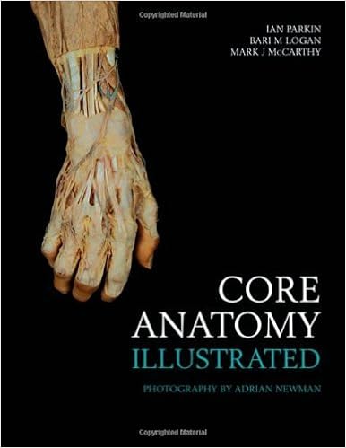
Middle Anatomy-Illustrated is a concise new atlas of human anatomy for clinical and allied wellbeing and fitness undergraduates, postgraduate trainees, and instructing employees. It offers the main anatomical wisdom precious for scientific perform, at a degree that's suitable with present middle curricula, both conventional or systems-based.Presenting superbly photographed dissections of remarkable readability, this succinct quantity includes seventy one easy-to-read double web page spreads. the mix of concise textual content at the left with annotated representation at the correct, deals a short, actual reference advisor to human anatomy.Written by way of Professor Ian Parkin, Mr. Bari Logan, and Mr. Mark McCarthy, who among them have over seventy five years event of training, analyzing and getting ready human anatomical fabric, center Anatomy-Illustrated covers the human physique, either female and male, actually from head to toe. The content material of the e-book has been rigorously chosen because the such a lot crucial 'end view' point of dissected anatomy that scientific, paramedical, and surgical practitioners needs to be acquainted with to perform correctly and successfully.
Read or Download Core Anatomy - Illustrated PDF
Similar medicine books

Oxford American Handbook of Disaster Medicine (Oxford American Handbooks in Medicine)
Mess ups are tough to control for plenty of purposes: the immediacy of the development, value of the development, loss of evidence-based practices, and the constrained usefulness of many constructed protocols. for this reason, combining educational ways with life like and useful strategies remains to be an underdeveloped point of catastrophe texts.
Taurine (2-aminoethanesulfonic acid) is an enigmatic compound abounding in animal tissues. it truly is current at particularly excessive concentrations in all electrically excitable tissues similar to mind, sensory organs, middle, and muscle, and in convinced endocrine glands. a few of its physiological features are already validated, for instance as a vital nutrient in the course of improvement and as a neuromodulator or osmolyte, however the mobile mechanisms are nonetheless usually an issue of conjecture.


- The New health care for profit : doctors and hospitals in a competitive environment
- Deja Review: Family Medicine (2nd Edition)
- Uterine Artery Embolization
- Diabetes und Schwangerschaft: Präventionen, Beratung, Betreuung vor, während und nach der Schwangerschaft
Extra resources for Core Anatomy - Illustrated
Example text
The internal carotid artery, surrounded by its plexus of sympathetic nerves, passes through the petrous temporal bone in the carotid canal (31), which opens in the skull immediately above the foramen lacerum. The internal jugular vein is formed in the jugular foramen (32), and cranial nerves IX, X and XI emerge from the foramen anterior to the vein. The occipital bone fuses with the body of the sphenoid anterior to the foramen magnum (33); behind the foramen it forms the posterior aspect of the cranium, which gives attachment to many small but powerful muscles that hold the head extended or rotate it at the atlanto-axial joint.
The spinal cord, closely covered by pia mater, is suspended in cerebrospinal fluid in the subarachnoid space. A flange of pia on each side sends fine denticulate ligaments to anchor the cord, via the arachnoid, to the overlying dural sac. These, and the filum terminale, a fibrous extension of the cord running all the way to the coccyx, prevent excessive movement of the cord. There is an epidural (potential) space (6) containing fat and a plexus of valveless veins between the dura and the bone, and ligaments of the vertebral canal.
32 Head and Neck Orbital skeleton, eyelids, conjunctiva The eyeballs are surrounded by muscles and supported by fat within the orbit. They must maintain their position and move in absolute synchrony, or double vision (diplopia) will ensue. Each bony orbit opens on the facial skull, bounded by the frontal bone (1), zygomatic bone (2) and maxilla (3). The orbit is cone-shaped with the apex passing backward and medially. This angulation is important when considering the actions of the orbital muscles.


