By Michael Chen, Thomas Pope, David Ott
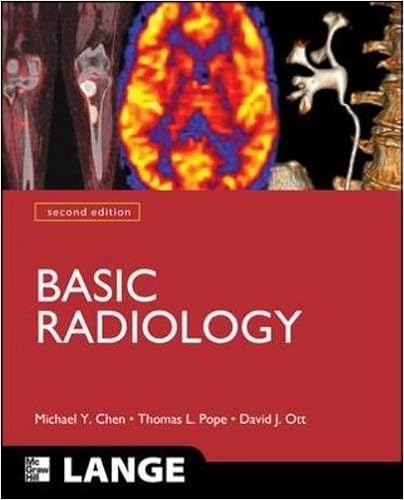
A well-illustrated, systems-based primer on studying radiologic imaging uncomplicated Radiology is the best and ultimate method for clinical scholars, citizens, and clinicians no longer focusing on radiologic imaging to benefit the necessities of diagnostic attempt choice, program, and interpretation. This relied on consultant is unequalled in its skill to coach you ways to choose and request the main acceptable imaging modality for a patient’s featuring signs and make yourself familiar with the commonest illnesses that present radiologic imaging can top evaluation. positive aspects: greater than 800 high quality pictures throughout all modalities A logical organ-system procedure constant bankruptcy presentation that comes with: ---Recap of contemporary advancements within the radiologic imaging of the organ procedure mentioned ---Description of ordinary anatomy ---Discussion of the main acceptable imaging process for comparing that organ procedure ---Questions and imaging workouts designed to reinforce your figuring out of key rules short record of advised readings and common references well timed bankruptcy describing some of the diagnostic imaging strategies at the moment on hand, together with traditional radiography, nuclear medication, ultrasonography, computed tomography, and magnetic resonance imaging a huge bankruptcy supplying an outline of the physics of radiation and its comparable organic results, ultrasound, and magnetic resonance imaging
Read Online or Download Basic Radiology, Second Edition (LANGE Clinical Medicine) PDF
Best radiology & nuclear medicine books
Textbook of Interventional Neurology
Endovascular intervention - utilizing medicine and units brought via catheters or microcatheters positioned into the blood vessels via a percutaneous method - has emerged as a comparatively new minimally invasive method of deal with cerebrovascular disorder and doubtless intracranial neoplasms. This textbook offers a complete assessment of rules pertinent to endovascular therapy of cerebrovascular illnesses and intracranial tumors, with an in depth description of strategies for those tactics and periprocedural administration suggestions.
Radiation Physics with Applications in Medicine and Biology
An advent to nuclear physics together with chapters on current and earlier purposes of nuclear medication, nuclear imagery, compartment thought, neutrons and different heavy debris, X-rays and Y-rays. contemporary details at the ideas of radiation defense can also be addressed.
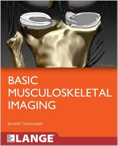
An entire introductory textual content to musculoskeletal imaging easy Musculoskeletal Imaging is an engagingly written, accomplished textbook that addresses the basic rules and strategies of common diagnostic and complicated musculoskeletal imaging. so one can be as clinically suitable as attainable, the textual content makes a speciality of the stipulations and approaches normally encountered in real-world perform, reminiscent of: top and decrease extremity trauma Axial skeletal trauma Arthritis and an infection Tumors Metabolic bone illnesses Bone infarct and osteochondrosis Shoulder, knee, backbone, elbow, wrist, hip, and ankle MRI additionally, you will locate authoritative assurance of: indicators in musculoskeletal imaging the main suggestions of utilizing diversified modalities in musculoskeletal imaging present advances in musculoskeletal scintigraphy The e-book is greater through fantastic figures and illustrations, together with a four-page full-color insert; "Pearls" that summarize must-know details; and a very good creation to musculoskeletal ultrasound through foreign specialists from France and Brazil.

Esophageal Cancer: Prevention, Diagnosis and Therapy
This publication stories the hot development made within the prevention, prognosis, and therapy of esophageal melanoma. Epidemiology, molecular biology, pathology, staging, and diagnosis are first mentioned. The radiologic review of esophageal melanoma and the function of endoscopy in analysis, staging, and administration are then defined.
- Pathology of the Pancreas: A Practical Approach
- Proton Therapy Physics
- Novel Trends in Brain Science: Brain Imaging, Learning and Memory, Stress and Fear, and Pain
Extra info for Basic Radiology, Second Edition (LANGE Clinical Medicine)
Example text
TECHNIQUES AND NORMAL ANATOMY A variety of techniques have been developed to evaluate the heart and great vessels (Table 3-1). In this section, we briefly describe the major tests used in imaging this system. ᮣ Conventional Radiographs The most common imaging test for evaluating the heart and great vessels is the chest radiograph, which consists of an upright posterior-to-anterior (PA) and left lateral (LAT) projections. The terms PA and left lateral refer to the direction the x-ray beam takes through the body before it reaches the 26 ᮡ PART 2 CHEST Table 3-1.
The catheter, introduced percutaneously into the thoracic aorta via the common femoral artery or placed into the ascending aorta at the time of surgery, should be positioned so that its tip is just distal to the origin of the left subclavian artery. The tip of the catheter has a small radiopaque marker so that this position can be ascertained on the chest radiograph (Figure 3-22). The major complications of the IAPB result from positioning of its tip proximal to the left subclavian artery, which may cause occlusion of the left subclavian vessel orifice, cerebral artery embolization, or aortic tear.
The anterior border of the cardiac shadow is composed primarily of the anterior wall of the right ventricle. Right ventricular enlargement may also encroach into the retrosternal clear space. The posterior margin of the cardiac silhouette is formed by the left atrium and left ventricle. Just posterior and inferior to the left ventricle is a linear soft-tissue shadow leading into the heart formed by the inferior vena cava (IVC). The left ventricular shadow should not project more than 2 cm posterior to the posterior border of the IVC.


