By Peter F. Semple
This useful sized pocket reference is designed to supply knowledgeable yet concise evaluation of the most important components of high blood pressure that might be of curiosity and relevance to clinicians. hugely illustrated in complete colour, with lucid textual content and tough self-assessment questions, the e-book could be of curiosity to citizens and different physicians fearful to replace their wisdom during this box.
Read or Download An Illustrated Pocketbook of Hypertension PDF
Similar cardiovascular books
Clinical Challenges in Hypertension II
This identify provides an leading edge new method of healing decision-making and gives solutions to a number questions that the busy clinician faces every day. a chain of discussions, chosen through professional participants, all the world over recognized specialists of their fields, provide their very own techniques for dealing with tough difficulties and provide insights into the worth or differently of varied therapy offerings in response to their very own adventure and the to be had proof.
Cardiovascular Magnetic Resonance: Established and Emerging Applications
An authoritative, up to date source at the frequently intimidating technical enigma of CMR, Cardiovascular Magnetic Resonance: demonstrated and rising purposes consolidates a large and becoming physique of data right into a unmarried balanced resource. This hugely illustrated textual content presents present and functional info on confirmed foundational functions of CMR.
Intracranial Hypertension (Neurology- Laboratory and Clinical Research Developments)
Intracranial high blood pressure (ICH) is the most typical reason for scientific deterioration and loss of life for neurological and neurosurgical sufferers. there are lots of motives of raised intracranial strain (ICP) and elevated ICP can produce intracranial high blood pressure syndromes. tracking of intracranial strain and advances in investigations of the principal worried procedure have ended in new recommendations and systemisations in intracranial high blood pressure.
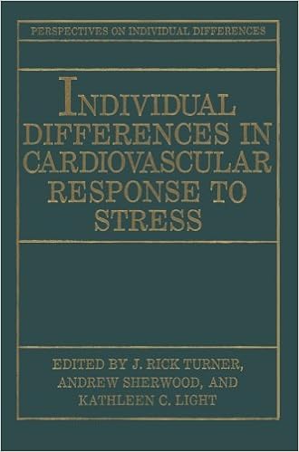
Individual Differences in Cardiovascular Response to Stress
Demonstrating that the significance and trend of cardiovascular reaction to emphasize varies markedly among contributors, this paintings discusses the mechanisms in which the cardiovascular process is mobilized in the course of rigidity, the determinants of person changes, and the pathophysiological methods in which responses to emphasize could lead on to heart problems.
- Rapid Review of ECG Interpretation
- Questions, Tricks, and Tips for the Echocardiography Boards
- ABC of Interventional Cardiology (ABC Series)
- Hypertensive Cardiovascular Disease: Pathophysiology and Treatment
- The Fifth Decade of Cardiac Pacing
Extra resources for An Illustrated Pocketbook of Hypertension
Example text
I ... ~,--- TIM E 0 V. C. G. RIGHT VENTRICULAR HYPERTROPHY This record shows regular sinus rhythm. 00, and the P axis is +15 degrees. The horizontal (JIS axis has a double transition, one between V2 and V3, the other at VII-V6. There is clearly excess anterior force. The horizontal T axis is about -110, the P axis is about +10 degrees. There is evidence of left and possible right atrial enlargement. The rightward and anterior QRS forces suggest right ventricular hypertrophy, and the T changes suggest right ventricular strain (and/or anterior ischemia).
0] SEC. /\..... STANDARD LEFT ATRIAL ENLARGEMENT 40 Courtesy III Dr. 10lIl14 Selvesl« IIJncho los Amigoi Hospili. -- I'~ ~ -----........ $. = ~ -'\~r SEQUENCE Of "YOCAIIOIAL DEPOLAl\llATlOfl I 10 SEC. OR LESS PEAKED IN LEAD II TEND. nihl Courtesy 01 Dr. ROIIIId S••••i1er (EXCEPT PUL EMPHYSEMA) PREDOM. 1\.. I~ II:::JC STANDARD RIGHT ATRIAL ENLARGEMENT Courtesy III Dr. ROIIII4 Selvesw IIJnchQ los Ami905 Hoipili' TIME RELATIONSHIPS ATRIAL ENLARGEMENT clinical explanation for the ST-T changes that occur with ischemia and "injury", and for the changes seen with hyper- and hypokalemia.
This difference is greatest during diastole, and is least just after the spike potential, when the cells are on the plateaus of their action potentials. This is why an "ST segment shift" is really a baseline shift. We also see that it is not telling us anything about the mystical word "injury", and we see no "current of injury". The EKG does not measure current. 41 It is simply a voltmeter. An ST segment shift therefore usually tells us that we have a significant group of cells that is working under severe ischemia or under adverse mechanical circumstances, and that the resting potentials of these cells are reduced compared to that of the normal cells.


